
The results of the first part of our work focused on evaluating the effectiveness of known RNA dimerization domains. In the course of this stage, we conducted a series of experiments to assess RNA–RNA dimerization, the possibility of forming double Cas complexes, and to measure the dissociation constants and functional activity of these systems in model eukaryotic cells.

Figure 1. Scheme of experiments in vivo.
During the in vivo tests, we used a dimerization stem-loop described in the study by Mutsuro-Aoki H., “Acquisition of Dual Ribozyme-Functions in Nonfunctional Short Hairpin RNAs through Kissing-Loop Interactions,” Life, 2022. The standard sgRNA scaffold was modified with RNA hairpins, and their activity was analyzed using several complementary methods. These modifications enabled the formation of RNA–RNA dimers and stabilized double complexes of dCas proteins on a model DNA molecule.

Figure 2. 10% TBE gel results of sgRNA structures modified with different hairpins, stained with SyBr Green (EMSA image).
As shown in the figure, the modification with the A-type hairpin (see sequence in Experiments) most actively promoted the formation of RNA–RNA pairs. This sequence was further analyzed for its potential to form dimers between dCas proteins.

Figure 3. Results of the 10% TBE gel for complexes of varying concentrations.
The obtained data indicate that the complexes successfully formed dimers with a model DNA molecule, confirming that this hairpin structure can be used to create stable dCas–RNA complexes.
In addition, the activity of this system was examined using Bio-Layer Interferometry (BLI).

Figure 4. Kissing-loop RNA–dual Cas protein–DNA and normal sgRNA–dual Cas protein–DNA combinations. Measurement of dissociation constants using BLI.
As shown in the figure, there was a noticeable stabilization of Cas complexes on the targeted DNA molecule compared to the standard guide RNA. This result demonstrated the potential of using structures such as kissing-loop RNA to form more stable double complexes. In subsequent stages, this structure was used to measure the activity of the complexes in in vivo models.

Figure 5. Bar graph showing the median GFP fluorescence intensity in K562 cells transfected with the modified CRISPR–dCas9 system.
Flow cytometry analysis of K562 cells showed no statistically significant decrease in GFP expression levels. Consequently, we decided to design a new system that would enable the generation of additional RNA–RNA interaction variants to improve the functional parameters of the complexes.
When selecting target sequences, we relied on the study by Yu-Chan Yang et al., “Permanent Inactivation of HBV Genomes by CRISPR/Cas9-Mediated Non-cleavage Base Editing,” Mol Ther Nucleic Acids, 2020. This work evaluated the activity of Cas proteins in different regions of the hepatitis B viral genome. After analyzing the results presented by the authors, we selected the most optimal and effective targets and applied these sequences in our experiments. The complete nucleotide sequences used are listed in Experiments.
At the second stage of our work, we focused on creating an efficient system for generating and selecting stable pairwise interactions between two modified sgRNAs.
A schematic representation of the experimental design is shown below.

Figure 6. General scheme of RNA–RNA interaction screening.
To achieve this goal, we designed and constructed a set of plasmids carrying different fragments of the genetic cassette required to obtain and select the most stable RNA–RNA dimers in dCas complexes.
The complete plasmid schematics and maps are provided in Experiments.
The system consisted of three functional plasmids:
Table 1. Expression Plasmids
| Plasmid Name | Description |
|---|---|
| P3sgRNAgen | Control plasmid |
| P3sgRNAp3p7 | Plasmid with a standard sgRNA scaffold |
| P3sgRNAp3p7n10 | Scaffold with 10 random nucleotides at the 3′ end |
| P3sgRNAp3p7n15 | Scaffold with 15 random nucleotides |
| P3sgRNAp3p7n20 | Scaffold with 20 random nucleotides |
Table 2. Sensor Plasmids
| Plasmid Name | Description |
|---|---|
| P1gP3gP7A | Plasmid with a promoter and two target sites (before and after screening) expressing TetR |
| P1gP3gP7B | Plasmid with two targets upstream of the promoter |
| P1gP3gP7R | Plasmid with two targets downstream of the promoter |
During the experiments, all three plasmid types were constructed and functionally tested. Cotransfection produced cells containing all plasmid combinations, which were then analyzed for clone viability and growth dynamics in the presence of tetracycline.

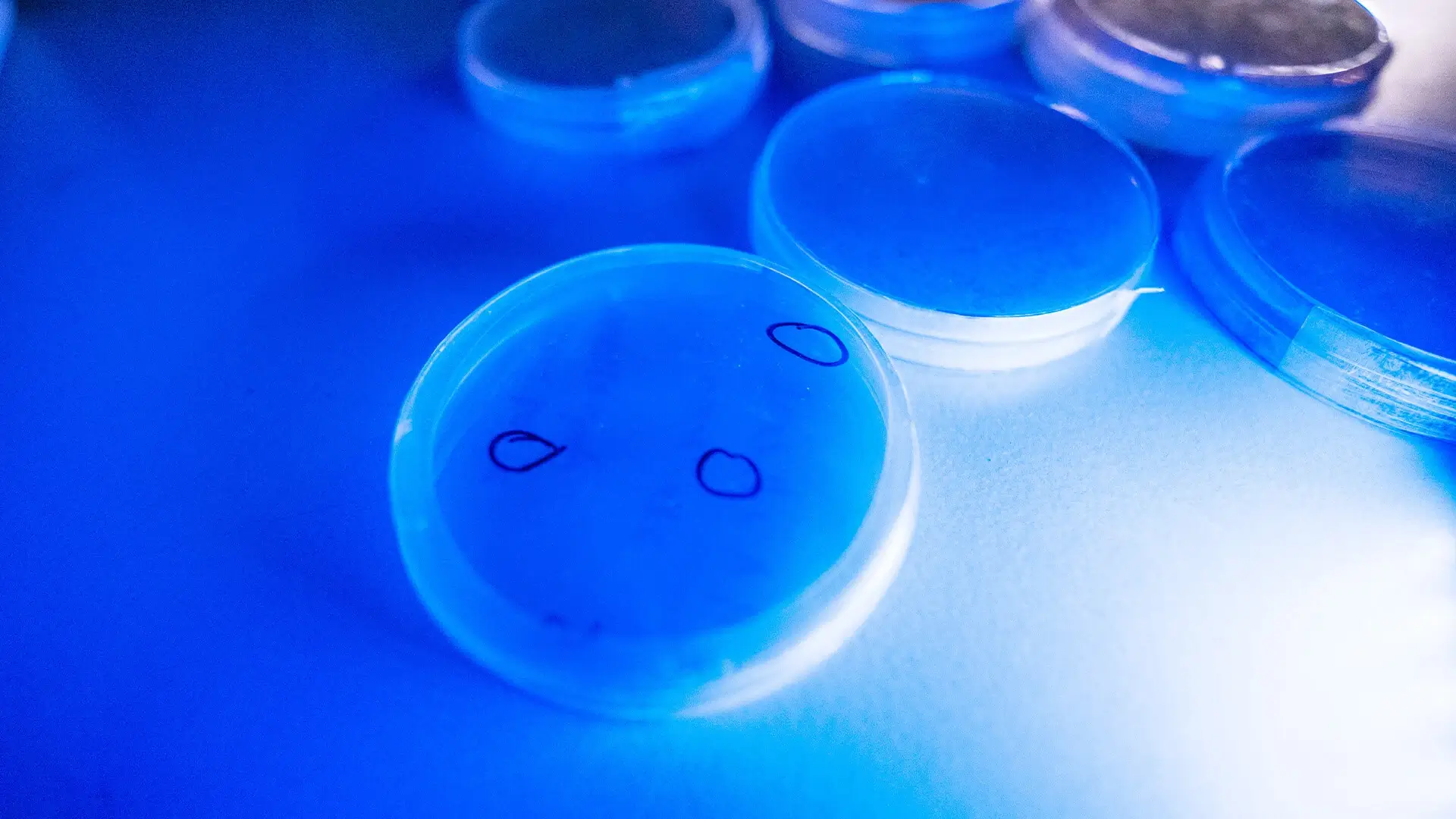
Figure 7. Growth of bacterial clones on tetracycline-containing plates. Tetracycline concentration increases from 10 ng/mL (top) to 100 µg/mL (bottom).
When analyzing the results, we observed an unexpected trend: as the concentration of tetracycline increased, cells carrying the tetracycline-resistance gene failed to survive at concentrations above 10 µg/mL. This finding indicated insufficient promoter activity in the sensor plasmid and revealed the need to modify the regulatory system for improved responsiveness.
Despite this limitation, our experiments confirmed high activity of double Cas complexes on a model DNA substrate, and we successfully established a functional three-plasmid system. Subsequent control tests using tetracycline further demonstrated that replacing the constitutive promoter with a stronger variant would significantly improve system efficiency.
Learn more in Experiments.